What We Can Now Say About the White Clots: From Molecular Insights to Scientific Truth
An Update for the Aware Community
Many dangers have emerged from gene injections imposed on the world since 2020, but those responsible for watching and acting have ignored them, claiming their evidence says everything is fine.
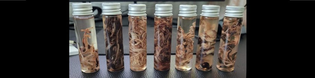
The science is becoming clearer on one thing though: the anomalous white fibrous casts being found too commonly in blood vessels are not ordinary clots. Two independent research teams have now published complementary findings that confirm this beyond reasonable doubt.
The Rapley trilogy of papers was published in late January 2026 on a preprint server. Now in peer review for journal publication, it provided the first comprehensive characterisation of these anomalous intravascular casts (AICs). The research was significantly funded by private citizens through NZDSOS, and conducted by New Zealand-based researchers Drs Bruce Rapley and Matt Shelton, using international labs on three continents.
The Santiago, Harrison et al paper follows in July 2026. Published in the International Journal of Innovative Research in Medical Sciences, their analysis including Raman spectroscopy study focused specifically on the protein structure of the casts.
Both teams reached the same fundamental conclusion: these are highly abnormal clots. And together, the two bodies of work provide a very coherent picture.
The Core Discovery: These Are A New Type of Clot
The central finding of the papers is that these fibrous casts are fundamentally different from expected pre-and post-death thrombi. They are not simply blood clots. They are massive misfolded protein aggregates that have undergone a specific, stage-dependent structural transformation.
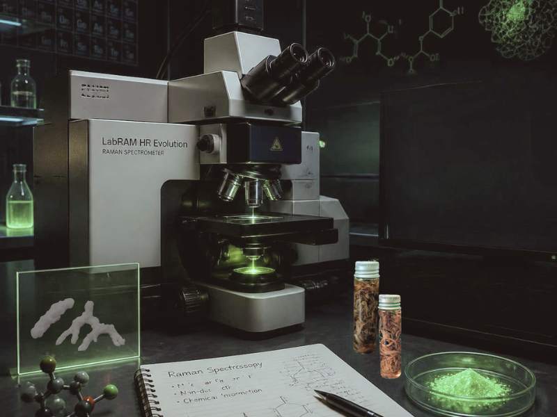
Raman micro-spectroscopy uses laser light to produce a unique chemical footprint of the specimen being analysed. At the HUN-REN Wigner Research Centre for Physics in Budapest, the Santiago team used Raman to identify that these casts contain areas enriched in beta-sheet structures. This is a molecular hallmark of amyloid, where proteins have lost their normal, functional shape and lock into dense, pathological fibres.
Dr Miklos Veres, one of the authors and a recognised world expert in Raman spectroscopy, described the transformation as follows:
“The proteins have not merely lost their native fold. They have adopted a new, thermodynamically stable architecture reinforced by backbone hydrogen bonding, aromatic packing, electrostatic phosphorylation contacts, and covalent disulfide bridges. This combination of stabilizing interactions is consistent with a persistent, self-assembling amyloidogenic scaffold rather than a reversible aggregation event.”
The Santiago team did not rely on Raman alone. They built a comprehensive evidence base:
Complementary Thioflavin T Fluorescence: ThT is a dye that is largely non-fluorescent in solution but lights up like a neon sign when it wedges itself into the grooves of amyloid fibrils. The green glow in their images confirms that the material is as near to amyloid as probably doesn’t matter.
Kjeldahl Protein Analysis: Confirmed a significant amount of crude protein (7.72%), proving these are fundamentally protein-based structures.
Ion-Exchange Chromatography: Profiled the specific amino acids, providing a molecular map consistent with stable, aggregated protein material.
The elevated proline and lysine in the amino acid analysis make sense in the context of blood vessel damage from immune attack.
If spike protein is damaging the arteries, the body will attempt to repair them. Embalmers and doctors have shown the clots often show multiple anchoring/originating points of attachment inside the vessels. Collagen is the scaffolding of many body tissues and repair requires proline and lysine. Indeed some integrative doctors in NZDSOS have long used these amino acids, along with vitamin C, in approaching cardio-vascular disease.
The presence of these amino acids in the casts may be a signature of that attempted repair process gone wrong.
The extremely low cysteine (0.01 m/m%), which contains sulphur, is also telling. Rapley and Shelton presumed that low sulphur indicated lower protein content. However, fibrinogen normally contains many cysteine residues that form disulphide bonds, and sulphur should show up in spectroscopy. The fact that the hydrolyzed fraction during spectroscopy shows so little cysteine could suggest that the disulphide bonds in the casts are not being broken down by acid hydrolysis – meaning they are unusually stable, possibly due to the amyloid-like structure protecting them – and therefore “invisible” to the analysis.
What We Can Now Say with High Confidence
1. These casts are structurally different from normal clots
Although this is clearly obvious to the naked eye, the Rapley morphology paper established that AICs are elastic, lumen-conforming, branched structures that form under active blood flow – in other words, during life. Yet they are devoid of intact red blood cells and platelets. Their rubber-like consistency and cohesive strength are highly incompatible with known pre- and post-mortem changes.
The difference was not subtle. The researchers compared AICs to clots usually produced before and after death and found them distinct from both.
2. They are biochemically abnormal
The elemental analysis revealed a bizarre chemical fingerprint: apparent sulphur depletion and relative phosphorus enrichment. This composition is incompatible with typical protein-dominant fibrin matrices and could not be explained by any conventional clotting mechanism.
The proteomic analysis then helped solve the protein puzzle. While the clots do contain fibrinogen (the building block of normal clots), the chains are in a highly abnormal ratio: approximately 1:7:3 for alpha:beta:gamma chains versus the normal 1:1:1.
Critically, plasminogen – the enzyme required to break down clots – was detected at extremely low abundance. This explains their stubborn persistence. This is reflected in what some clinicians and their patients are telling us – the normal clot-buster drugs are not working in many cases.
3. Structurally they seem like amyloid. This is a very bad thing.
The Harrison team’s Raman spectroscopy confirmed that these casts contain beta-sheet structures, a molecular hallmark of amyloidosis. But they do acknowledge that final absolute confirmation of the fibrous clots being amyloid relies on even higher tech testing not yet done. They have other work in progress, and some provisional findings which may be even more alarming.
4. They show a maturation pathway
Both teams observed evidence of staging. The Rapley team noted the presence of partial Lines of Zahn, indicating formation under active blood flow. The Santiago-Harrison group identified two distinct spectroscopic states in samples: S1 (alpha-helix dominant, early changes/native-like protein structure) and S2 (beta-sheet enriched, advanced/mature amyloid). These were samples from 2 different people, with multiple runs of analysis carried out.
This suggests these structures are not static. Whilst looking clearly abnormal, they can progress from a relatively ‘typical’ state toward a highly ordered, stabilised amyloid-like form.
5. They seem structured to persist
The combination of findings paints a coherent picture of persistence:
- The abnormal fibrinogen chain ratio (1:7:3) suggests a fundamentally different fibrin architecture
- The profound plasminogen deficiency (0.1283%) means the body cannot break them down
- The amyloid beta-sheet structure provides physical stability
- The disulphide bridges and phosphorylation contacts lock the structure in place
Researcher Dr Bruce Rapley stated: “This is not just a big blood clot. This is a fundamentally different architecture. The profound deficiency in plasminogen is like building a structure impervious to future demolition – it’s designed to persist”.
What This Means for the Broader Picture
The two studies together provide a mechanistically coherent explanation for persistent vascular obstruction, impaired oxygen delivery, and a broad spectrum of acute and chronic symptoms reflecting organ dysfunction.
They also point to urgent questions. The Rapley paper explicitly notes: “If spike protein were demonstrated to provoke anomalous intravascular casts, this would raise serious implications not only for covid pathophysiology but also for genetic platforms that induce sustained host manufacture of spike protein, making it imperative that this potential association be rigorously investigated”.
What We Still Can’t Say for Sure
We don’t know for absolute certain that lab-engineered covid-19 or its jabs, and the spike protein in common with both, have caused this blood disease. But until epidemiological studies are done which prove otherwise, this seems the case. As research continues the spike protein looks more like a main culprit.
How common are they? Embalmers surveys are very alarming, 20 to 30 % of all embalmings according to another peer-reviewed paper by Daniel Santiago, along with the authors of the clotastrophe blog. But how many people are wandering around unknowing if their own bodies are harbouring these things? Again, formal acknowledgement is required before studies can start.
Are they pathogenic (disease or death-causing)? As long as most specialists remain in denial and refuse to investigate, common sense must provide the answer. Restricting blood flow in blood vessels, big and small, is never going to end well.
Are they related to the microscopic amyloid versions described in pre-covid times, and in spike protein syndromes since 2020? Most likely. The only difference is size, and the site and nature of the trigger for production. This is a very useful review of the work that builds the bridge between pre-covid and the subsequent time of spikeopathy.
Why Happens Next?
The two streams of research represent independent lines of evidence pointing to the same conclusion: these are a novel, persistent, pathologically abnormal form of intravascular material. So there is amyloid-like damaged protein forming in blood which will block blood vessels of all sizes.
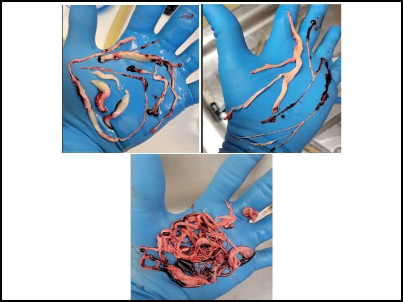
This is simply awful in its implications and must be confronted, but the denial of this reality has been the real crisis. We have told everyone relevant about these clots, and will continue to do so. There is no of lack of evidence. The photos of these twisted worm-like clots (or like a bundle of spaghetti, as a local embalmer just told us) are shocking enough, let alone handling the real thing.
Yes, we can see that the world’s senior bureaucrats and elected politicians are already serving a shadow global government. Ex UK prime minister Liz Truss has just admitted it. But is the refusal to look at the clots also due to simple human psychology? Denial is a powerful weapon against psychological pain. And the willfully blind will stay that way until enough of us force them to look.
As usual with denial, though, these people are hurting themselves just as badly as the rest of us. There may be solutions for the (very likely) spike protein-induced disease of blood vessels and the blood itself, as discussed in this expert webinar hosted by Dr Philip McMillan – one of very few medical consultants willing to discuss this new disease. But scaling up any treatments for the truly gigantic number of people probably affected will require official recognition. Clearly, we need some new officials.